Dr Saba Vakili MD, Histopathology Specialty Registrar·
Breast cancer is the most common cancer in women worldwide and remains one of the leading causes of cancer-related deaths. According to the World Health Organisation, more than 2 million new cases of breast cancer are diagnosed globally each year, affecting women across all regions and populations.
Despite these numbers, advances in screening, diagnosis, and treatment have significantly improved outcomes. Today, many women diagnosed with breast cancer can expect successful treatment and long-term survival, particularly when the disease is detected early.
This is where breast cancer screening plays a vital role.
What Is Breast Cancer Screening?
Breast cancer screening is the process of examining individuals who have no symptoms of breast cancer to identify disease at an early stage.
The goal is simple: to detect cancer before it becomes large enough to cause symptoms or spread beyond the breast.
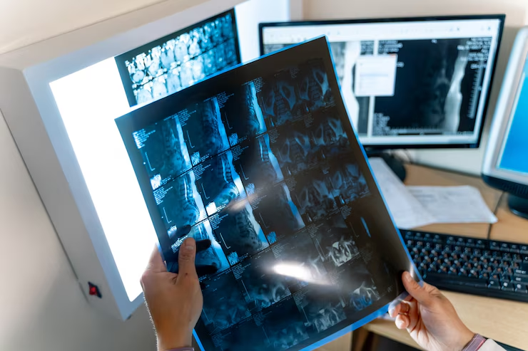
Most screening programmes use mammography, a specialised low-dose X-ray examination of the breast. Mammograms can reveal abnormalities that may be too small to be felt during self-examination or routine clinical examination.
By identifying cancers earlier, screening can improve treatment options and reduce deaths from breast cancer.
Why Is Early Detection Important?
The stage at which breast cancer is diagnosed is one of the most important factors influencing treatment and survival. When breast cancer is found early:
- Treatment is often less extensive
- Breast-conserving surgery may be possible
- The likelihood of cure is higher
- Survival rates are significantly improved
Many breast cancers detected through screening are found before any symptoms develop, providing an opportunity for earlier intervention and better outcomes.
Breast Screening Around the World
Many countries have established organised breast cancer screening programmes, although eligibility criteria and screening intervals vary.
Examples include:
- The United Kingdom, which offers routine mammographic screening through the NHS Breast Screening Programme.
- Several European countries that provide population-based screening every two to three years.
- Canada and Australia, which operate national or regional screening programmes.
- The United States, where screening recommendations vary between professional organisations but generally encourage regular mammography for women at average risk beginning in midlife.
In addition to organised national programmes, many healthcare systems offer opportunistic screening, where mammograms are arranged during routine healthcare visits.
Regardless of the specific approach, the underlying principle remains the same: detecting breast cancer before symptoms appear.
The NHS Breast Screening Programme: An Example
One of the largest and most established screening programmes is the NHS Breast Screening Programme in the United Kingdom.
Women registered with a general practitioner are routinely invited for screening mammography every three years between the ages of 50 and 71 years. Individuals outside this age range may still be eligible in certain circumstances or may request screening depending on local policies.
What Happens During a Mammogram?
A mammogram is a relatively quick procedure performed by trained healthcare professionals.
During the examination:
- Each breast is positioned on the mammography machine
- Gentle compression is applied for a few seconds
- X-ray images are taken from different angles
The procedure usually takes less than 30 minutes
Although some women experience temporary discomfort during compression, the examination is generally well tolerated.
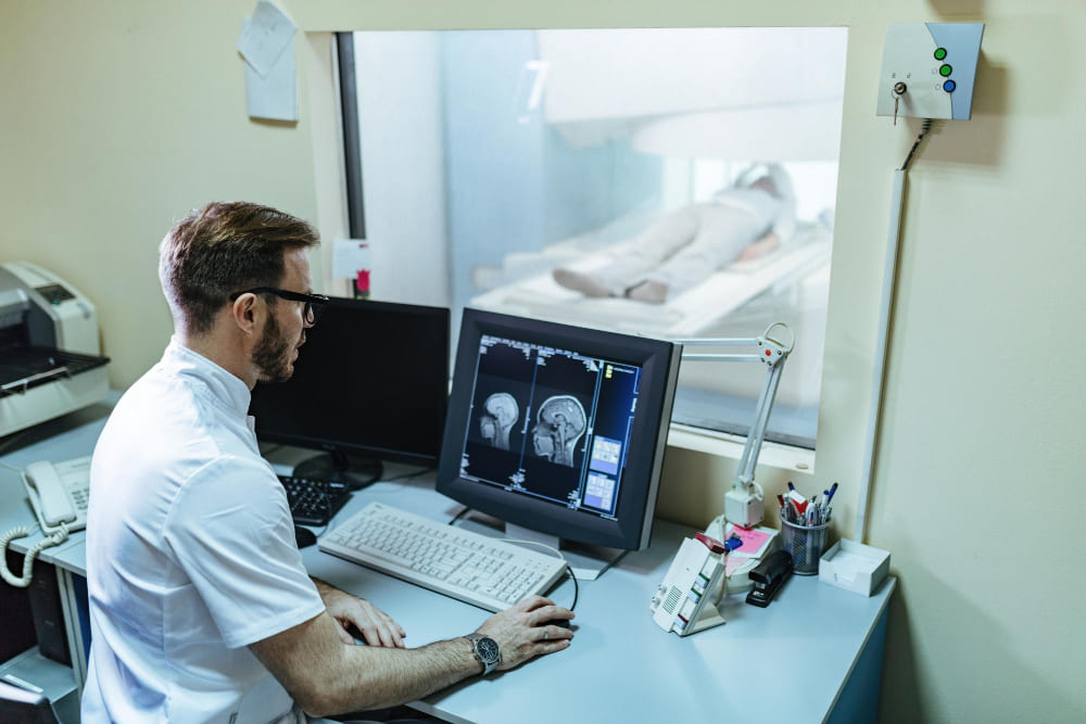
The images are then reviewed by specialist breast radiologists.
What Happens If an Abnormality Is Found?
Being recalled after a screening mammogram does not necessarily mean that cancer is present.
Many abnormalities detected during screening are ultimately found to be benign conditions such as cysts, fibroadenomas, or other non-cancerous breast changes.
However, further investigation is essential to determine the nature of the finding.
This assessment is typically performed using a well-established process known as the triple assessment.
Understanding the Triple Assessment
Triple assessment is considered the gold standard for investigating breast abnormalities. It combines three complementary approaches that together provide a highly accurate diagnosis.
1. Clinical Assessment
The first component is a clinical evaluation by a breast specialist.
This includes:
- Reviewing symptoms and medical history
- Discussing family history and risk factors
- Physical examination of the breasts
- Examination of the lymph nodes, particularly in the armpit region
2. Radiological Assessment
The second component involves imaging studies.
Depending on age and clinical circumstances, imaging may include:
- Ultrasound
- Breast MRI in selected cases
Radiological assessment allows specialists to evaluate the size, location, and characteristics of any abnormality and helps guide subsequent investigations.
3. Pathological Assessment
The third component is pathology, which provides the definitive diagnosis.
If an area appears suspicious, a biopsy may be performed. A small sample of tissue is removed using a needle and examined under the microscope by specialist pathologists.
Pathology can determine:
- Whether a lesion is benign or malignant
- The specific type of breast cancer
- Tumour grade
- Important biomarkers that help guide treatment decisions
Because pathology directly examines tissue, it is often considered the final step in confirming a diagnosis.
Why Is Triple Assessment So Effective?
Each component contributes unique information:
- Clinical examination assesses what can be seen and felt.
- Imaging evaluates the internal structure of the breast.
- Pathology examines the tissue at a microscopic level.
When the findings from all three assessments agree, diagnostic accuracy is extremely high. This multidisciplinary approach ensures patients receive the most appropriate diagnosis and treatment recommendations.
When Should You Seek Medical Advice?
Breast screening is designed for people who do not have any symptoms. However, it is important to remember that breast cancer can develop between screening appointments, and some cancers occur in individuals who are not yet eligible for routine screening.
You should consult a healthcare professional if you notice any new or unusual changes in your breasts, including:
- A new lump or thickening in the breast or armpit
- Changes in the size or shape of the breast
- Dimpling, puckering, or redness of the skin
- Changes to the nipple, including inversion (turning inward)
- Unexplained nipple discharge, particularly if blood-stained
- Persistent breast or nipple pain affecting one area
- A rash or crusting around the nipple
- Swelling in the armpit or around the collarbone
Most breast changes are not caused by cancer, but it is important to have any new symptoms assessed by a healthcare professional.
Do not wait for your next screening appointment if you develop symptoms. Screening is intended to detect cancer before symptoms appear, while any new breast change should be investigated promptly regardless of your age or when your last mammogram was performed.
Key Take-Home Messages
- Breast cancer is the most common cancer affecting women worldwide.
- Early detection significantly improves treatment options and survival.
- Mammography is the most widely used breast cancer screening tool.
- Many countries operate organised breast screening programmes, while others provide screening through routine healthcare services.
- The NHS Breast Screening Programme is an example of a population-based screening system designed to detect cancer before symptoms appear.
- Any abnormality identified during screening is typically investigated using the gold-standard triple assessment approach: clinical examination, radiology, and pathology.
- Screening saves lives, but individuals should still seek medical advice promptly if they notice any breast changes or symptoms.